Helpful Nanoparticles Inhibiting and Focused on Conveyance Applications
Benita Sultana*, Nibret Hoekstra
Massachusetts General Hospital Cancer Center, 55 Fruit St, Yawkey 9E, Boston, MA, 02114, USA
Correspondence to: Benita Sultana, Massachusetts General Hospital Cancer Center, 55 Fruit St, Yawkey 9E, Boston, MA, 02114, USA. E-mail: benita_sultana@harvard.edu
Received: February 11, 2023; Accepted: February 27, 2023; Published: March 06, 2023
Citation: Sultana B, Hoekstra N. Helpful Nanoparticles Inhibiting and Focused on Conveyance Applications. Arch Virol Infect Dis. 2023;1(1):1-7.
Copyright: © 2023 Sultana B. This is an open-access article distributed under the terms of the Creative Commons Attribution License, which permits unrestricted use, distribution, and reproduction in any medium, provided the original author and source are credited.
ABSTRACT
Nanoparticles are wide used as remedies for disorders for a protracted time. They're 10-9 m specks of gear that may be found each naturally and synthesized within the laboratory with metal and nonmetal materials during this study, gold nanoparticles (AuNPs) were synthesized exploitation the turn reduction methodology, and therefore the thirty-five nm size of the nanoparticles resolve employing a UV-Vis photometer at 525 nm wavelength. The synthesized nanoparticles were additional studied on MCF-7 carcinoma cells to grasp however numerous genes square measure expressed within the induction of cell death in signal transduction pathways. The results obtained from the antineoplastic activity of the gold nanoparticles showed roughly ninetieth inhibition of cell growth once seventy-two hours of treatment. Western blot analysis incontestable the downregulation of p44/42 MAPK (ERK1/2) macromolecule thanks to gold nanoparticle treatment. Moreover, reverse transcription-polymerase chain reaction (RT-PCR) analysis of apoptotic genes discovered the upregulation of the p53 growth suppressor, Bax, and caspase-9. The results assembled from this study additional indicates that p44/42 MAPK, p53, proteinase nine and bax play a serious role within the mechanism of cell death within the MCF-7 carcinoma cells.
KEYWORDS
AuNPs; Gold nanoparticles; MCF-7; Apoptosis; Sequence expressions; Cancer.
ABBREVIATIONS
MCF: Michigan Cancer Foundation; AuNPs: Gold Nanoparticles; ATCC: yankee sort Culture Collection; FBS: craniate Bovine Serum; MTS: 3-(4,5-dimethylthiazol-2-yl)-5-(3-carboxymethyl phenyl)-2-(4-sulfophenyl)- 2H-tetrazolium); PBS: Phosphate Buffered Saline; M-PER: class macromolecule Extraction Reagent; BCA: Bicinchoninic acid; TBST: Tris Buffered Saline and Tween 20; MAPK: Mitogen-Activated macromolecule Kinase; immune serum globulin HRP: immune gamma globulin endocrine cathartic Peptide; GAPDH: aldehyde 3-Phosphate Dehydrogenase
INTRODUCTION
Nanotechnology has become an awfully rampant analysis topic over the past years starting from in vitro to in vivo clinical analysis exploitation numerous metal and nonmetal particles targeting cancer cells and tumors [1]. One in all the established blessings of the nanoparticles is its size and its potential to be programmed to specifically target organelles or tumors [2]. Gold is popularly called a component on the tabular array with Associate in Nursing number of +3 and +1. it's Associate in Nursing number of seventy-nine Associate in Nursing a mass of 197 amu. It's a transition metal and is solid at temperature. Gold is usually present however may be synthesized, that is rare. tho' gold has been used for a moment in dentistry|odontology} medicine, physical science, and physical science engineering, alittle size (nanoparticle) has been accepted by multiple researchers as a probable agent for cancer remedy in biomedicine [3]. A well-liked methodology for gold nanoparticle synthesis could be a reduction reaction of tetra chloroauric acid (HAuCl4) by turn normally known as the Turkeivich methodology. different reducing agents like plants as illustrated by Raghunandan et al. and Geetha et al. and fungi [4-6] are used also. Researchers have additionally rumored however the potential sizes and shapes of synthesized gold nanoparticles [7-10].
MATERIAL AND METHODS
Cell Culture
Cells were purchased from ATCC (American sort Culture Collection) and maintained in five-hitter DMSO at -80°C till use. For culture, cells were maintained in RPMI 1460 Medium (Gibco, CA) enriched with 100% FBS (Fetal Bovine Serum) and 1 Chronicles antibiotic/ antifungal agent at 37°C with five-hitter greenhouse emission during a humidified atmosphere. Cells were plated at five × 104 cells/well to get a pattern of growth for additional studies.
Synthesis of Gold Nanoparticles
The nanoparticles were synthesized employing a modification of the Turkevich methodology by Cesbron et al. [11]. Briefly, 0.01 g of HAuCl4 was more to fifty metric capacity unit of heated water. 1 Chronicles of Na turn (Fisher Scientific) was then more to the heated stirring resolution. a discount reaction was detected once the answer changed into a wine color. the answer was pure employing a zero.2 two filter, shielded from lightweight and hold on at 4°C.
Treatment of Cells
MCF-7 cells were seeded at three × a hundred and five cells per plate, allowed to connect for twenty-four hours and incubated during a humidified atmosphere at 37°C with five-hitter greenhouse emission. The cells were treated with zero, 5, 10, fifteen and twenty μg/ml of AuNPs in culture media. Cells were counted exploitation the improved Neubauer hemocytometer chamber for five consecutive days.
Cell Viability by MTS Assay
The MCF-7 cells were plated at a density of one × 104 cells per well during a ninety-six well plate and allowed to connect. Thereafter, the cells were treated with AuNPs at zero, 5, 10, fifteen and twenty μg/ml for seventy-two hours. Manufacturer’s (Promega) directions were followed. Briefly, twenty of MTS resolution was more to every well and incubated for four hours. The plates were browsing at 492 nm employing a SkanIt plate reader. the info obtained were statistically analyzed.
Western Blot Analysis
The MCF-7 cells (5 × 10⁵) were incubated in petri dishes and allowed to connect for twenty-four hours. The cells were treated with similar concentrations as explicit higher than of AuNPs for 3 consecutive days, trypsin zed exploitation EDTA-Trypsin (Gibco), washed with PBS and hold on at -20°C. Macromolecule was extracted from the cells exploitation M-PER (Mammalian macromolecule Extraction Reagent) (with proteinase Inhibitor) and was quantified exploitation the BCA assay kit (Thermo Scientific). The samples (20 μg) of macromolecule to be run were mixed with lysis buffer and loading buffer (dye) and place into the ten SDS-PAGE (Nupage 100% Bis-Tris Gel, Invitrogen). The gel was run at 160V so transferred to the cellulose ester membrane (Bio-rad, 0.45μm) at 120V. The membrane was blocked exploitation five-hitter blotting grade milk powder resolution in 1x TBST for Associate in Nursing hour and washed within the same buffer. Thereafter, the membrane was soaked long in p44/42 MAPK (1:1000) primary protein. The membrane was washed thrice with 1x TBST Associate in Nursingd soaked during a secondary protein (Goat anti-Mouse immune serum globulin HRP 1:2000) for an hour. The membrane was then stripped and immersed in GAPDH primary protein with a corresponding secondary protein. once repetitive laundry, the blot was soaked in luminescence (Clarity Western ECL Substrate, Biorad) resolution. pictures were pictured exploitation the ChemiDoc Imaging System and computer code (Biorad).
Real-Time Quantitative Enzyme Chain Reaction
The MCF-7 cells (5 × 10⁵) were plated in petri dishes and allowed to connect for twenty-four hours. Cells were treated for seventy-two hours with similar concentrations as explicit higher than of gold nanoparticles. polymer was extracted from the cells exploitation the Aurum Total polymer mini Kit (Biorad). Briefly, the cells were lysed exploitation lysis resolution supplemented with β-methcarpenol, repetitive laundry with hour plant product, high and low stringency resolution and DNase resolution was wont to extract the polymer during a binding column. Finally, the extraction resolution at 70ºC was wont to collect the polymer extract for every sample. For the reverse transcription method, the polymer model was incubated for Associate in Nursing hour at 42°C with iScript Reverse Transcription Supermix (Biorad) for deoxyribonucleic acid synthesis. employing a SsoAdvanced Universal SYBR inexperienced Supermix (Biorad), and five primers for every sample (p53, Caspase 9, BaCl2, Bax, and β-actin). PCR plate (Hard-Shell PCR Plates 96-well, thin-wall, Biorad) was started and run forty cycles at 98°C (30 secs), 95°C (15 secs) and 60°C (25 secs) to get Ct values employing a CFX96 instrument and computer code (Biorad).
STATISTICAL ANALYSIS
Statistical information for the reverse transcription-polymerase chain reaction was obtained in relevance β-actin. the strategy of calculation was obtained from Kannan [12]. The distinction between the Ct values of ablated and treated samples were obtained for all of the targeted genes (p53, caspase-9, bcl2, bax, and β-actin). The distinction between these 2 fresh obtained values was calculated to the negative power of 2, for effort the relative fold modification.
RESULTS
Characterization and Purification of Gold Nanoparticles
The synthesized AuNPs were determined employing a UV-Vis photometer measured at 525 nm wavelength. tho' there square measure studies that have characterized gold nanoparticles by matter surface assimilation [12], the AuNPs during this analysis were classified consistent with the Cytodiagnostics Charts [13] and a mathematical study conducted by Rahman. The nanoparticle size was calculable to be regarding thirty-five nm size Associate in Nursingd an approximate concentration of regarding fifty μg/ml. the answer was pure employing a zero.2 two filter. Figure 1 showed the spectrum of the synthesized AuNPs from the UV-Vis photometer.
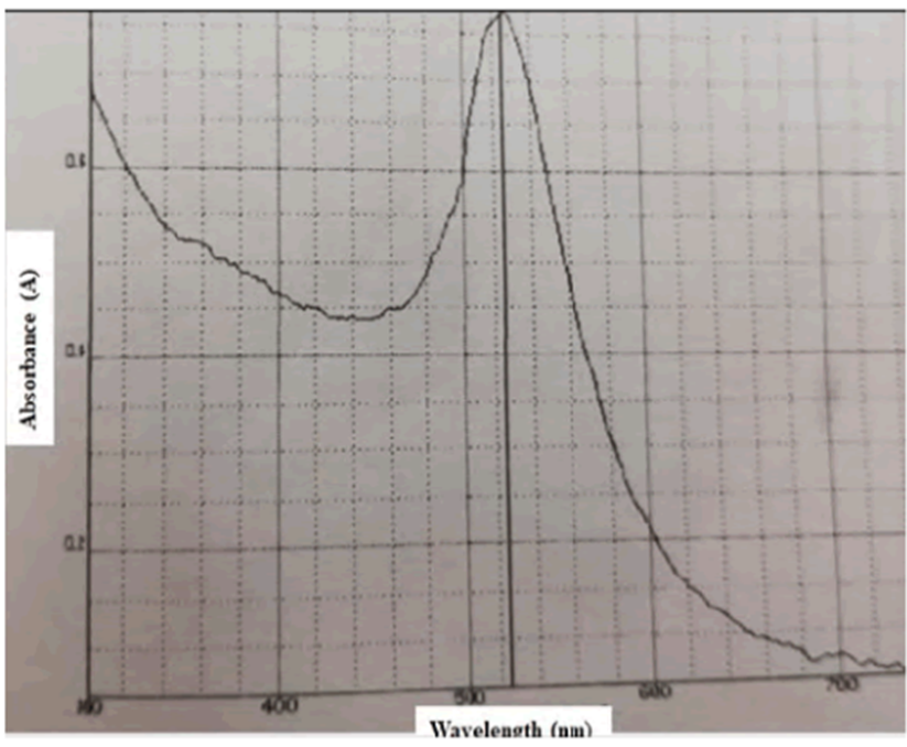
Figure 1: Characterization of synthesized gold nanoparticles. This figure shows the results of the UV-Vis Spectrophotometer.
Effects of Gold Nanoparticles on the Viability of MCF-7 Cells
Cells were treated with zero, 5, 10, fifteen and twenty μg/ml of AuNPs. the info was collected daily for five days to grasp the results of AuNPs treatment on this cell line. Figure 2 showed viability of the cells at completely different concentrations of AuNPs for five consecutive days. As shown in Figure 2, the cell proliferation was small as concentration of AuNPs was inflated. At twenty μg/ml of AuNP concentration, the cell viability was reduced by over five hundredth once twenty-four hours of treatment.
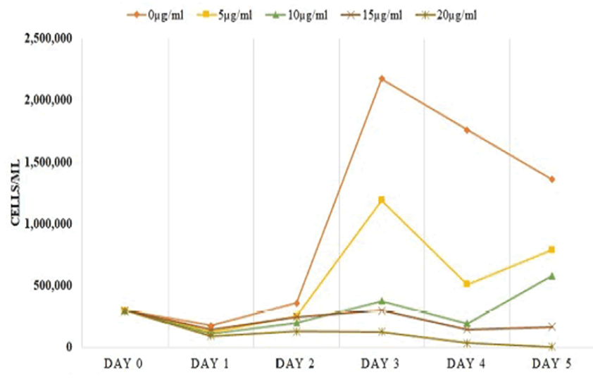
Figure 2: Effects of gold nanoparticles on the viability of MCF- 7 cells. MCF-7 cells were treated with 5, 10, 15 and 20 µg/ml concentrations of gold nanoparticles.
Inhibition of Proliferation Shown by MTS Assay
To measure the cytotoxic effects of AuNPs, the cells were treated at numerous concentrations of AuNPs and therefore the information was retrieved once seventy-two hours of treatment. Figure 3 showed that thanks to the rise in AuNPs concentration, there was a proportional increase in inhibition of cells consistent with absorbance. Compared with the management cells, the Figure 3 additionally showed thirty-five, fifty and ninetieth inhibition of the cells at the concentrations of five, ten and fifteen μg/ml of AuNPs, severally.
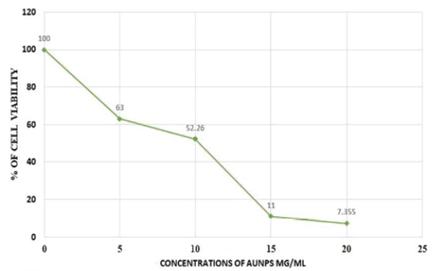
Figure 3: Cytotoxicity assay of gold nanoparticles on MCF-7 cells using MTS assay.
Downregulation of p44/42 MAPK by Western Blot Analysis
To depict the results of the AuNPs on the phosphorylated proteins p44/42 within the MAPK pathway, western blotting was performed there was a downregulation of p44/42 once treatment with AuNPs indicating the inhibition of the phosphorylation that is correlative with the impediment of neoplastic cell growth. This figure additional showed a decrease in information measure by five hundredth compared to the management, also because the consistency of the information measure of GAPDH as a house-keeping macromolecule.
Activation of p53, caspase-9, and Bax Indicated by RT-qPCR
RT-PCR offers quantitative data regarding the expression of genes. The quantitative information of the organic phenomenon by RT-PCR. applied mathematics computations were meted out meted out to get double delta Ct for every of the averages of the targeted sequence samples. The 9-fold upregulation of the p53 sequence, that could be a growth suppressor, suggesting that there was a major increase in p53 expression once treatment with AuNPs. The expression of caspase-9 sequence was inflated 3-fold, this indicating that there was important caspase-mediated necrobiosis thanks to the treatment with AuNPs. However, bax and bcl-2, each apoptotic regulator, were expressed to ~2.5-fold and ~0.6-fold, severally. The Bax seems to be considerably upregulated, bcl-2 expressions were appeared to be insignificant.
DISCUSSION
Nanoparticles don't seem to be novelle. Over the past decades there has been a high rise within the use of various types of nanoparticles. In 2012, Co chemical compound nanoparticles were rumored to induce cell death in Jurkat and K cell lines in vitro studies. Aptamer tagged paclitaxel loaded poly lactic-co-glycolic acid nanoparticles were shown to decrease viability in GI-1 cancer cells. Also, Rajeeth et al. disclosed that synthesized oxide nanoparticles inflated the expression of p53 considerably in MCF-7 cells once analysis through western blotting. Though, solely gold nanoparticles square measure employed in this study, there looks to be a powerful correlation within the activities of nanoparticles as remedies for cancer.
Most of the recent studies dole out have combined the gold nanoparticles with either light-weight or different biological substances. in a very study dole out by Bhowmik et al. wherever the results of gold nanoparticles with a poisonous substance obtained from AN cobra on human lymphocytes and leukemic cells showed that there was iatrogenic programmed cell death in leukemic cells. Vedantam et al. reported that synthesized gold nanoparticles had a big impact on prostatic adenocarcinoma cells (DU-145) once 2-3 days. They additionally indicated the results of combined sugar D-mannan (Mn) with gold nanoparticles explaining that, there was doubly the maximum amount uptake than simply gold nanoparticles on prostatic adenocarcinoma cells. Peptides have additionally been conjugated with gold nanoparticles by Kalmodia et al. to be tested on Y79 malignant tumor cells (RCB1645). They stipulated that each peptide indicated dose-dependent inhibition of reactive chemical element species and additionally showing there was additional uptake once the gold nanoparticles were conjugated with the peptides. in a very study wherever grapes were wont to synthesize gold nanoparticles recommended that the plasma of the neoplastic cell (HBL- 100) was affected negatively and programmed cell death was investigated. Though the gold nanoparticles during this study weren't combined nor increased, they incontestable important effects on the MCF- seven cells. Therefore, it's that is crucial to focus on a number of the approaches that are taken victimization gold nanoparticles.
Several studies reported the activities of gold nanoparticles on a molecular side over the past years. These were targeting however gold nanoparticles may be answerable for reducing proliferation because of targeting organelles like mitochondria, endoplasmic reticulum, nucleus, and also the Golgi apparatus, lysosomes, proteasomes, etc. Epidermis protein has additionally been conjugated with gold nanoparticles to look at effects in vitro on the A549 carcinoma cell line. it absolutely was reported that the EGF conjugated AuNPs entered the cell’s living substance. Another study reported that a smaller size (1.9 nm) of gold nanoparticles suspended in sterile water has been reported to induce programmed cell death in DU-145 cell line once expressions of apoptotic agents like caspase-9 and cleaved PARP were analyzed by western blotting. This study solely targeted on the expressions of some sign proteins within the MCF-7 cell line treated with turn mixture gold nanoparticle that created the requirement for our study to account for the expression of different proteins. A closely connected analysis reported the expressions of most genes targeted during this study. In 2012, Selim et al. [10] reported that once treatment of MCF-7 cells with gold nanoparticles, the neoplasm gene p53, caspase-9, caspase-3, and Bax were considerably upregulated whereas bcl2 was downregulated. Our study additionally illustrates however p53, caspase-9, Bax, BCL-2 and p44/42 manifest in MCF-7 human breast carcinoma cells once treatment with gold nanoparticles. Therefore, it will be inferred that the rise of p53, Bax, and caspase-9 and downregulation of p44/42 conducts a substantial half on the mechanism of inhibition by gold nanoparticles.
CONCLUSION
In conclusion, this study confirmed that the viability of MCF-7 cells was attenuated with a rise within the concentration of AuNPs. The investigations were dole out to check the results of the cells’ signal transduction pathway once treatment with mixture gold nanoparticles. These results disclosed iatrogenic programmed cell death and inhibition of MCF-7 cells proliferation through the cistron expressions of p53, caspase-9, Bax and p44/42. This study encourages more analysis in vivo and prospective clinical trials victimization AuNPs as a metastatic tumor drug.
REFERENCES
1. Chauhan A, Zubair S, Tufail S, Sherwani A, Sajid M, et al. Fungus-mediated biological synthesis of gold nanoparticles: Potential in detection of liver cancer. Int J Nanomedicine. 2011;6:2305.
2. Guo J, Rahme K, He Y, Li LL, Holmes JD, et al. (2017) Gold nanoparticles enlighten the future of cancer theranostics. Int J Nanomedicine 12: 6131-6152.
3. Khanyile S, Masamba P, Oyinloye BE, Mbatha LS, Kappo AP (2019) Current biochemical applications and future prospects of chlorotoxin in cancer diagnostics and therapeutics. Adv Pharm Bull 9: 510-520.
4. Jain S, Hirst DG, Sullivan JMO (2011) Gold nanoparticles as novel agents for cancer therapy. Br J Radiol 85: 101-113.
Selim ME, Hendi AA (2012) Gold nanoparticles induce apoptosis in mcf-7 human breast cancer cells.Asian Pac J Cancer Prev 13: 1617-1620.
5. Cesbron Y, Shaheen U, Free P, Lévy R (2015) TAT and HA2 facilitate cellular uptake of gold nanoparticles but do not lead to cytosolic localisation.Plos One 10: e0121683.
6. https://bitesizebio.com/24894/4-easy-steps-to-analyze-your-qpcr-data-using-double-delta-ct-analysis/
7. https://www.cytodiagnostics.com/store/pc/Introduction-to-Gold-Nanoparticle-Characterization-d3.htm
8. Rahman S (2016) Size and concentration analysis of gold nanoparticles with ultraviolet-visible spectroscopy.Undergraduate Journal of Mathematical Modeling: One+Two: 7.
9. Chattopadhyay S, Chakraborty SP, Laha D, Baral R, Pramanik P, et al. (2012) Surface-modified cobalt oxide nanoparticles: New opportunities for anti-cancer drug development.Cancer Nanotechnol 3:13-23.
10. Aravind A, Varghese SH, Veeranarayanan S, Mathew A, Nagaoka Y, et al. (2012) Aptamer-labeled PLGA nanoparticles for targeting cancer cells.Cancer Nanotechnol 3: 1-12.
11. Rejeeth C, Kannan S, Muthuchelian K (2012) Development of in vitro gene delivery system using ORMOSIL nanoparticle: Analysis of p53 gene expression in cultured breast cancer cell (MCF-7).Cancer Nanotechnol 3: 55-63.
12. Bhowmik T, Saha PP, Dasgupta A, Gomes A (2013) Antileukemic potential of PEGylated gold nanoparticle conjugated with protein toxin (NKCT1) isolated from Indian cobra (Naja kaouthia) venom.Cancer Nanotechnol 4: 39-55.
13. Vedantam P, Huang G, Tzeng TR (2013) Size-dependent cellular toxicity and uptake of commercial colloidal gold nanoparticles in DU-145 cells.Cancer Nanotechnol 4: 13-20.